Infographics & Publications
Pelvic Matters
Outreach and Awareness Zine | Contributor
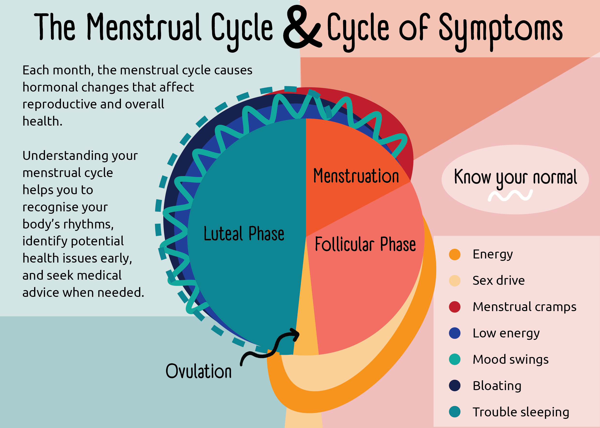
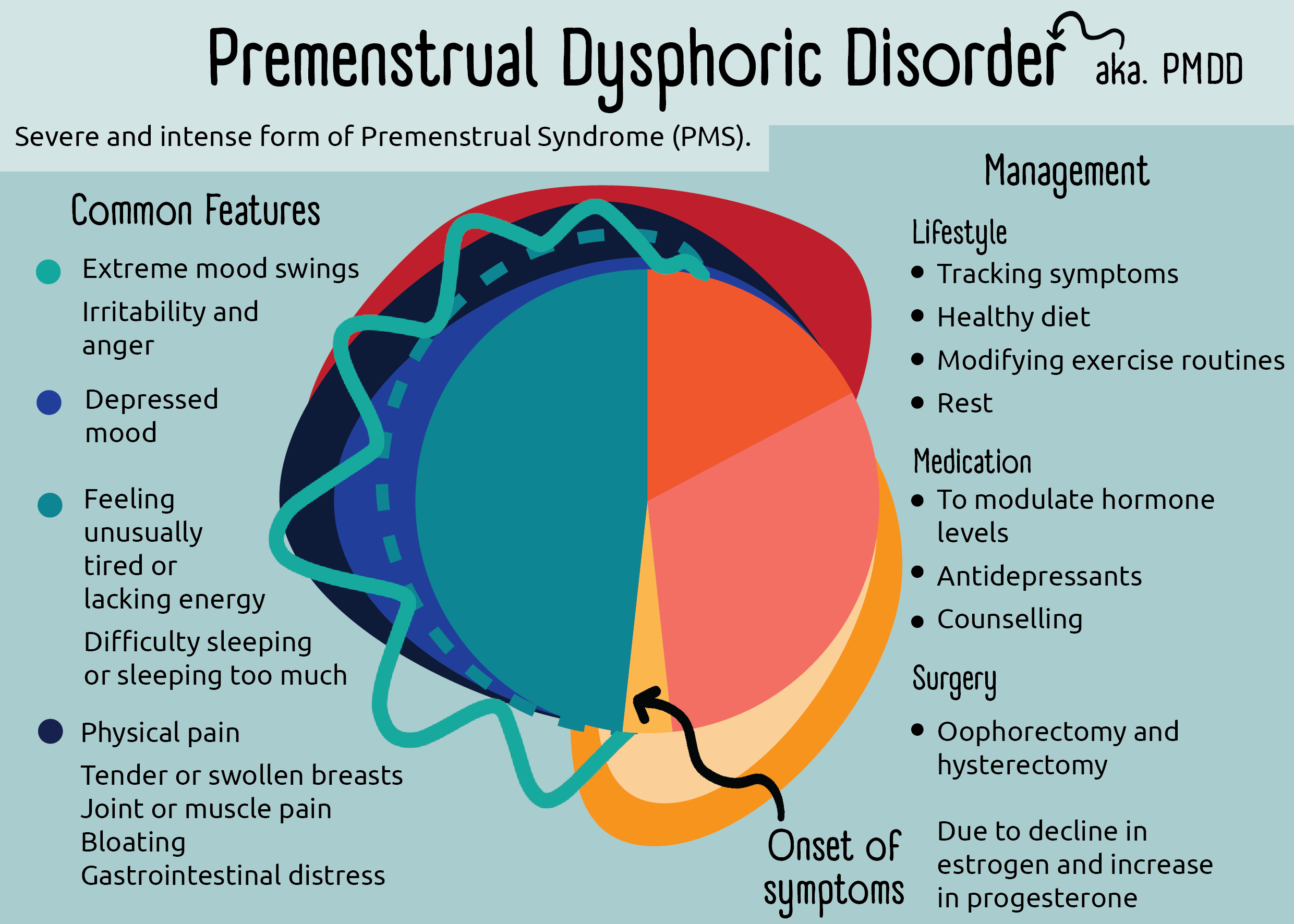
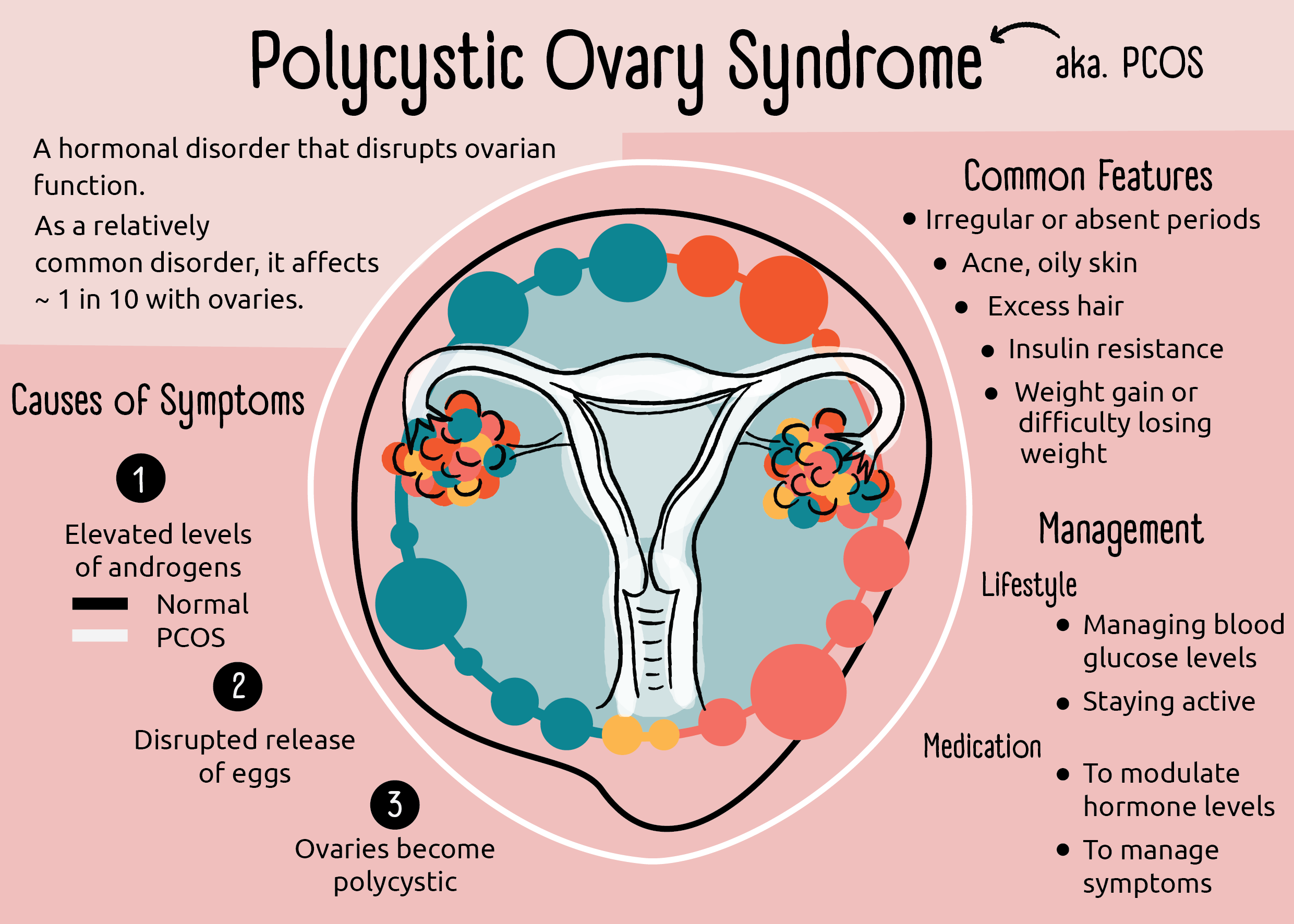
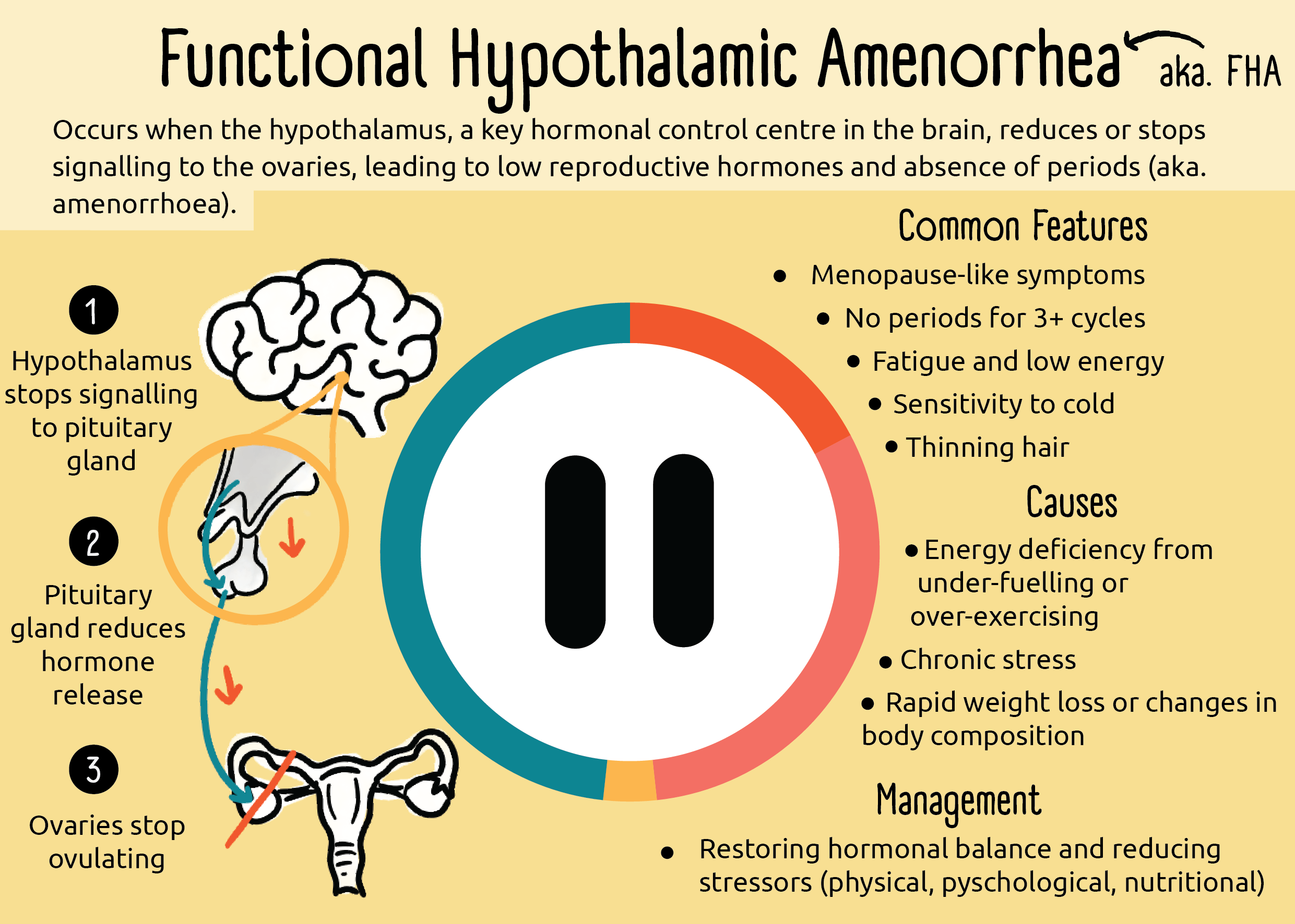
Created by early-career researchers at the Wellcome Genome Campus, Pelvic Matters is a pocket-size booklet that distils peer-reviewed science into clear illustrations and concise text. Inside you’ll find essential facts on female reproductive anatomy, the menstrual cycle, contraception, menopause and conditions such as endometriosis, polycystic ovary syndrome, and vulvodynia, all in a format that is easy to carry, read and share.
Why Science Needs Art
Perspective Paper | Author
Thesis Figures
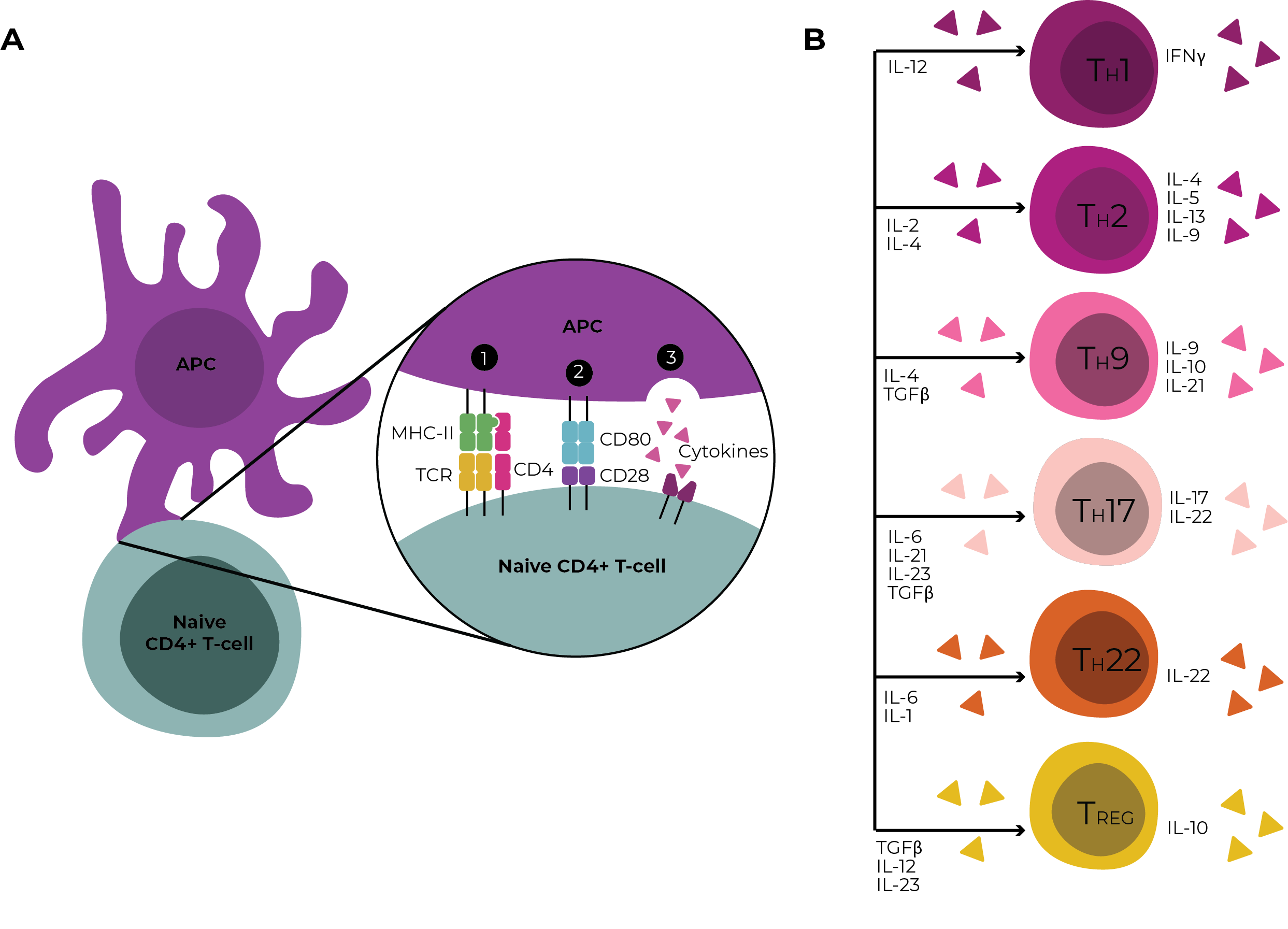
Introduction | Author
Figure 1.2: Three-signal model of CD4+ T-cell activation and CD4+ T-helper subset polarisation. A) Schematic of the interaction between a naive CD4+ T-cell and APC, illustrating the three signals required for full T-cell activation: (1) antigen recognition via TCR binding to peptide–MHC, (2) costimulatory signalling through CD28 engagement with CD80/CD86 and (3) cytokine signalling from the surrounding microenvironment. B) Overview of cytokine conditions that drive differentiation into selected T-helper (Th) subsets. The panel shows cytokines required for polarisation (left) and signature cytokines typically produced by each subset (right). APC= Antigen-presenting cell, MHC-II = Major histocompatibility complex class II, TCR = T-cell receptor, IL = Interleukin, TGF= Transforming growth factor.
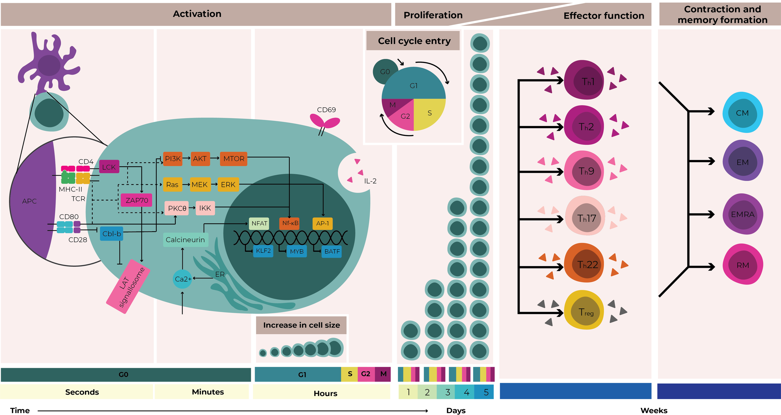
Figure 1.3: Temporal dynamics of CD4+ T-cell activation, proliferation and differentiation. Simplified schematic illustrating key molecular and cellular events following T-cell activation. From left to right: an antigen-presenting cell (APC) delivers TCR and CD28 costimulatory signals to a naive CD4+ T-cell, initiating intracellular signalling cascades. These cascades involve the activation of LCK, ZAP70 and the LAT signalosome. Costimulation also inhibits Cbl-b, an E3 ubiquitin ligase that, if active, targets LAT components for degradation and suppresses PI3K signalling. Downstream of these early events, transcription factors NFAT, NF-κB and AP-1 are activated, while transcriptional repressors of activation, including KLF2, MYB, and BATF are downregulated. Within the first few hours, early activation markers such as CD69 are upregulated and IL-2 is produced and secreted. mTOR signalling promotes metabolic reprogramming and cellular growth, enabling entry into the cell cycle and the onset of proliferation. As proliferation progresses, cytokine cues from the environment guide polarisation into distinct effector T-helper (Th) subsets. Over the course of one to two weeks, the expanded effector population contracts, giving rise to long-lived memory T-cell subsets: central memory (CM), effector memory (EM), effector memory re-expressing CD45RA (EMRA), and tissue-resident memory (RM) cells. An approximate cell cycle phase and activation timeline is indicated along the bottom of the figure. APC= Antigen-presenting cell, MHC-II = Major histocompatibility complex class II, TCR = T-cell receptor, IL = Interleukin.